Video of living cells captured by a microscope can reveal a lot of information about cell characteristics and behavior, but parsing that information from hundreds of frames showing thousands of cells would be incredibly time-consuming to do manually. Computer vision technology, such as the kind used for facial recognition, offers a means of analyzing many images at a high speed and can reduce the issues of subjective human bias and human error. Engineers at the Universidad Carlos III de Madrid (UC3M) have now developed a fully automatic computer vision system specifically designed to characterize the behavior of cells traveling through vessels within living tissue.
The new system is driven by a deep neural network that can be trained to recognize and distinguish between cells in a 3D spatial context within a blood vessel. After detecting and segmenting the cells the system can extract features such as shape, size, motion and position of individual cells relative to the blood vessel over time, tracking all of this data for up to thousands of cells in a tiny fraction of the time it would take a person to record manually. The software is also equipped with collision detection capabilities so the trajectory of individual cells can be accurately tracked even as the cells bump into one another, and the system can be used to classify cells into different groups based on their behavior patterns.
The system, titled Automatic Cell Migration Examination (ACME), was used by the researchers to analyze 4D (3D space plus time) intravital microscopy images of neutrophils in mouse blood vessels, showing how the neutrophils behaved differently during inflammatory processes. Analysis using ACME also allowed the researchers to identify that mice treated with an inhibitor of the FGR protein showed less pathogenic migration behavior of their neutrophils than untreated control mice, suggesting that FGR could play a role in cardiovascular disease. The ACME software has the ability to be trained using different datasets involving different cell types other than neutrophils, making it a flexible system with the possibility to be applied to many different research areas. This research was published recently in the journal Medical Image Analysis.
“It is not feasible to keep an expert biologist segmenting and tracking cells on video for months. On the other hand, to provide an approximate idea (because it depends on the number of cells and 3D volume depth), our system only takes 15 minutes to analyse a 5-minute video,” said paper co-author Ivan González Díaz.
The team is now using ACME to study other types of cells, including T cells and dendritic cells in cancerous tissues. This work was conducted in collaboration with researchers from the National Centre for Cardiovascular Research in Madrid, the Vithas Foundation, the University of Castilla-La Mancha, the Singapore Agency for Science, Technology and Research (ASTAR) and Harvard University.
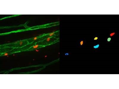
Photo: Neutrophil segmentation using the proposed system with ACME software. The segmentation is 3D, but an accumulated 2D version is shown. On the left, original microscopy image: blood vessels (green) and neutrophils (red). On the right, automatic segmentation using ACME (one colour for each cell). Credit: UC3M