Advanced X-ray imaging techniques allow researchers to explore the nanostructures of materials such as those used in microchips and batteries, but this type of imaging has traditionally been limited to the use of monochromatic X-rays. While imaging technologies that utilize visible light have for centuries benefited from the use of achromatic lenses, which allow multiple wavelengths to be focused on a single point, no such optical elements have been available for use with X-ray methods, and high-resolution X-ray microscopy often requires the use of expensive, high-brightness synchrotron sources to provide beams with enough intensity to achieve a clear image. Now, a team of researchers at the Paul Sherrer Institute (PSI) have developed a novel achromatic lens for X-rays, opening up the possibility of new compact imaging systems for materials science and R&D applications.
Chromatic aberration in visible imaging technologies can be resolved with the use of achromatic doublet lenses consisting of two different materials with high and low dispersion that together focus two different wavelengths into focus on the same plane. For X-rays, there is currently no pair of materials with the necessary optical properties to have the same effect, says physicist Christian David, head of the X-Ray Optics and Applications research group at PSI’s Laboratory for X-ray Nanoscience and Technologies. So, in order to design a novel achromatic X-ray lens, the team instead combined a focusing diffractive element and a defocusing refractive element to correct the chromatic aberration. For the diffractive element, a nickel Fresnel zone plate (FZP) was fabricated via electron-beam lithography and nickel electroplating, and for the refractive element, a novel structure consisting of four stacked paraboloids was 3D-printed using two-photon polymerization lithography.
The newly developed lens was tested using an X-ray beamline at the Swiss Light Source synchrotron at PSI. The researchers performed scanning transmission X-ray microscopy and ptychography experiments at a wide range of X-ray energies using the achromatic lens as a focusing optic, and imaged the same samples using a conventional FZP instead of the achromat. The achromatic lens allowed for much higher image quality at changing energies than the FZP, for which images became significantly blurred at different energies. With the 3D-printed refractive element designed by the researchers acting as a corrector for the chromatic behavior of the FZP, sharp X-ray images could be achieved over a wide range of wavelengths. This research was published in Nature Communications.
“Industry would like to have much faster response loops in their R&D processes. Our achromatic X-ray lens will help enormously with this: It will enable compact X-ray microscopes that industrial companies can operate on their own premises.”
PSI is now working with its spinoff company XRnanotech to market the new lens, and plans to connect with companies that specialize in building X-ray microscopy facilities on the laboratory scale. The technology could improve access to high-resolution microscopy capabilities for advanced materials research and industrial R&D without being restricted by the limited availability of high-power synchrotron sources.
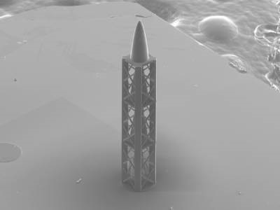
Photo: A microstructure created by a 3D printer: the innovative refractive structure developed by PSI scientists and which combined with a diffractive element, results in an achromatic X-ray lens, is almost a millimetre long (or high, as shown in the photo). Turned on its end, it resembles a miniature rocket. It was created by a 3D printer using a special type of polymer. This image of the structure was captured using a scanning electron microscope. Credit: Paul Sherrer Institute/Umat Sanli