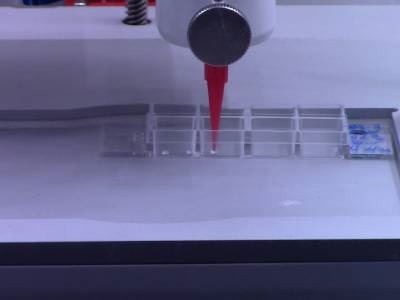
Plant cells are bioprinted by a 3D printer. Credit: Lisa Van den Broeck, NC State University
In nature, cells interact within a three-dimensional microenvironment, in which function and communication can be affected by positional and mechanical cues. It is difficult to reproducibly form such 3D microenvironments in the laboratory using current conventional culturing methods, but bioprinting – the 3D printing of living cells – offers a promising new avenue for studying cellular function and signaling. A research team from North Carolina State University recently demonstrated a 3D bioprinting method for plant cells that showed high cell viability, opening up new possibilities for high-throughput and reproducible studies of cells in 3D microenvironments.
The 3D bioprinting method is mechanically similar to methods used for 3D printing of other materials such as plastics. However, some optimizations were made to the bioprinting system, such as the inclusion of an ultraviolet filter to maintain a sterile environment, and the use of multiple print heads to print multiple bioinks simultaneously, explained first author Lisa Van den Broeck. The bioinks were designed with specific proportions of protoplasts, nutrients, growth hormones and agarose, and cells from both the model plant Arabidopsis thaliana and soybeans were printed for the study. Agarose was selected as an optimal scaffolding, as it forms a liquid during printing and solidifies after printing, and the compositions of the inks were designed to mimic a natural environment where cells would communicate as they would in soil, explained co-corresponding author Ross Sozzani.
Experiments showed that more than half of the 3D bioprinted cells were viable after printing and divided over time to form small colonies known as microcalli. The viability of the cells was similar to that of manually pipetted cells, with the printing enabling much more reproducible, faster and more precise deposition of cells. The researchers also bioprinted individual cells to test whether they could regenerate; in these experiments, more than 40% of individual soybean embryonic cells remained viable two weeks after bioprinting and also divided over time to form microcalli. Finally, the team studied the cellular identity of the bioprinted cells.
“We found that bioprinted cells can take on the identity of stem cells; they divide and grow and express different genes,” said Van den Broeck. “When you bioprint, you print a whole population of cell types. We were able to examine the genes expressed by individual cells after 3D bioprinting to understand any changes in cell identity.”
This study was published in Science Advances. The researchers plan to continue their work studying cellular communication after 3D bioprinting, including at the single-cell level. The technique could potentially be used in the future to study cellular regeneration in crop plants, said Sozzani.