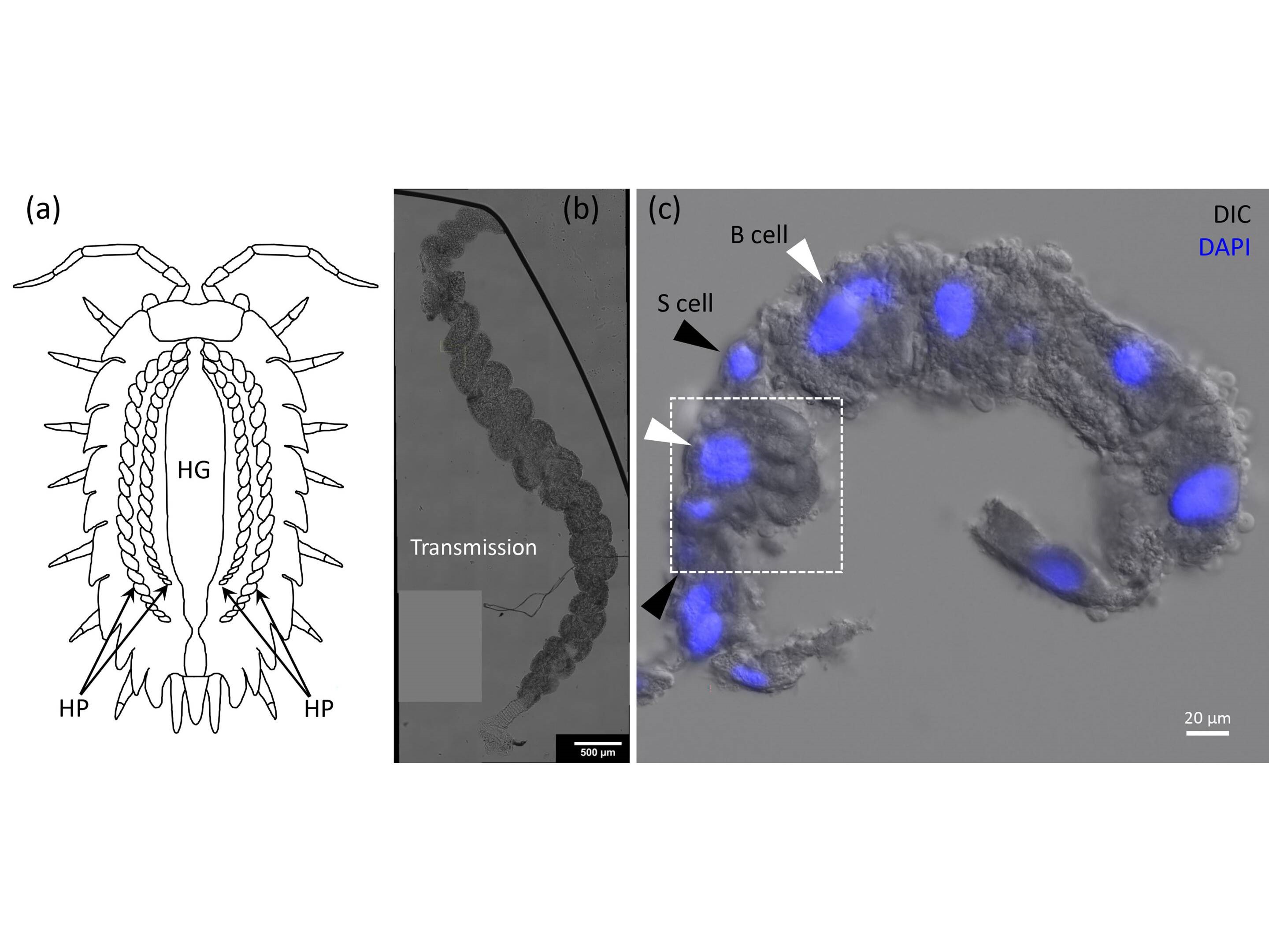
(a) Cartoon of a woodlouse depicting the hepatopancreas (HP) and the hind gut (HG). (b) Transmission overview of a single HP tubule, showing the helical structure. (c) Section from a HP tubule with the nuclei fluorescently labeled in blue. Credit: Iestyn Pope, Nuno G.C. Ferreira, Peter Kille, Wolfgang Langbein, and Paola Borri
Metal nanoparticles have the potential to leach from industrial sites, contaminating the environment and potentially impacting nearby wildlife. However, the true impact of metal pollution on organisms is not well understood due to a lack of methods to visualize metal nanoparticles at high resolution within living tissues. Researchers from Cardiff University have now developed a new microscopy method that could potentially be used to better understand the fate of metals ingested by organisms, using the unique properties of gold nanoparticles and woodlouse to their advantage.
Oniscus asellus, a small terrestrial isopod commonly known as woodlice, roly-polies or pill bugs, are useful for studying environmental metal contamination due to their ability to process metals using a specialized digestive organ called the hepatopancreas. The method designed by the researchers is based on four-wave mixing microscopy, in which three pulsed lasers of different frequencies interact with a material to produce a fourth frequency that can reveal highly detailed three-dimensional information about the material. In this case, the materials studied were gold nanoparticles ingested by the woodlice and processed in the hepatopancreas. Gold nanoparticles are especially useful in biological research due to their biocompatibility and photostability, noted corresponding author Wolfgang Langbein. The team attempted to use four-wave mixing microscopy to selectively locate the gold nanoparticles within the organ’s 3D cellular environment by tuning the laser pulses based on the localized surface plasmon resonance of the particles.
The researchers found that the technique enabled the nanoparticles to be visualized with subcellular resolution despite the fact that imaging intact organ tissues such as the hepatopancreas is typically challenging due to their high light scattering properties. The method even enabled single gold nanoparticles, 10 nm in radius, to be visualized within the complex, multicellular organ. The imaging method was relatively fast, at 0.1-1 ms per pixel, and used low excitation powers of less than 100 μW, meaning the method could potentially be used for live cell imaging. The experiments allowed the researchers to observe differences in the accumulation of gold within different specific cell types in the hepatopancreas. This study was published in Applied Physics Letters.
“By precisely pinpointing the fate of individual gold nanoparticles in the hepatopancreas of woodlice, we can gain a better understanding of how these organisms sequester and respond to metals ingested from the environment,” said Langbein. “Tracking this metal within these organisms is the first step enabling further study to determine, for example, if gold is collected within specific cells, or if it can interfere with the metabolism in high doses.”
The team believes their four-wave mixing microscopy-based technique could lay the groundwork for studying how metals are processed in the organs of environmentally-relevant organisms, and could be used in the future to locate other types of metal nanoparticles, and in different species such as fish larvae and human cell cultures.