University of Kentucky researchers have developed a simple method to observe metabolic shifts in cancer cells which could be an effective tool for future cancer research. Metabolic shifts are of growing research interest to further our understanding of how tumors adapt to, and thrive after, therapies are administered.
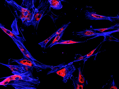
During their work, which is published in the journal Biophotonics Discovery, the researchers developed a novel microscopy technique which combines fluorescence microscopy and imaging software to observe and analyze metabolic changes in individual cancer cells. By employing advanced imaging techniques these shifts could be observed without the need for expensive equipment or destructive testing.
While focusing on head and neck squamous cell carcinoma (HNSCC) the team found that radiation treatments led to significant metabolic shifts in the cells, particularly the activation of HIF-1α which helps the cells adapt to low oxygen levels. Additionally, they found that one specific HNSCC cell line exhibited much higher levels of HIF-1α expression, suggesting a shift toward radioresistance. They also found that by reducing HIF-1α expression, a cells radioresistance could be reversed.
“The study demonstrates the functional flexibility of our novel optical approach to report the key metabolic changes of radioresistant and radiosensitive HNSCC under therapeutics stress thereby revealing the role of metabolism reprogramming in the development of resistance to cancer therapeutics,” said Senior author Caigang Zhu.
“This work was motivated by the practical barriers for the access to expensive metabolic tools we met in the past for tumor metabolism studies,” Zhu added. “Our demonstrations and results are exciting, as we now have a cost-effective approach to study cell metabolism at single-cell level with minimal expertise requirement.”