Three-dimensional fluorescence imaging of neurons in Layer 5 of the cerebral cortex, captured by two-photon microscopy in mice expressing a fluorescent protein (Thy1-EYFP-H). Credit: Shigenori Inagaki and Takeshi Imai, Kyushu University
If you’re sitting in a laboratory right now, especially a life science one, odds are there is a bottle of bovine serum albumin (BSA) somewhere nearby. The reagent is widely used in protein quantification, as a blocking agent in immunoassays, as a stabilizer, as a cell culture supplement and more. Its stability, low cost and high purity make it ideal for these applications.
Researchers at Kyushu University in Japan have just—accidentally—discovered another function it is ideal for: turning a living brain “transparent” without disturbing function.
In a paper just published, the Japanese team introduces a new reagent called SeeDB-Live that uses albumin to clear tissue while preserving cellular function. This allows scientists to see deeper, brighter structures in both brain slices in a dish and living mice, reaching neural activity that was previously out of sight.
“This is the first time tissue clearing has been achieved without altering its biology,” said Takeshi Imai, professor at Kyushu University and the study’s senior author, who has been working on this for over 10 years. After developing reagents for fixed tissue in 2013 and 2016, Imai concluded it would be impossible to create something similar for live tissue clearing.
“That question came to me about 100 times, and each time I answered ‘impossible,’” he recalled. “But 10 years later, here we are. When something seems unachievable, if you keep thinking about it, you may eventually find a way.”
Fail, fail, succeed
Complex functions like memory and thought arise from real-time communication between cells deep in the brain. Although slices preserve some activity, understanding normal brain dynamics requires imaging the living brain—which is incredibly difficult. Making the opaque brain transparent is one solution, but that has not been done in a living brain while preserving biological activity—until now.
Through systematic experiments, Imai’s team found that living cells become most transparent when the refractive index of the extracellular solution is adjusted to 1.36–1.37.
With a precise target in hand, the team needed a non-toxic way to reach it while maintaining osmotic balance, so that cells neither swell nor shrink. They previously tried natural substances such as sugar, but these required high concentrations that increased osmotic pressure and dehydrated cells.
As osmotic pressure depends on the number of molecules, the team turned to large spherical polymers. Their greater size means fewer are required to raise the refractive index, which adjusts optical performance without overwhelming the cells. However, despite screening nearly 100 compounds, they could not find a solution.
Then, late one night, Assistant Professor Shigenori Inagaki grabbed a bottle of bovine serum albumin (BSA) to try. To his surprise, BSA showed the lowest osmotic pressure at the desired refractive index.
By adding albumin to the culture medium to match the refractive index inside cells, Inagaki, Imai and team then developed a live-tissue clearing solution, which they named SeeDB-Live.
How it works
According to the study published in Nature Methods, SeeDB-Live renders mouse brain slices transparent within 1 hour of immersion. When combined with a calcium indicator, the normal neuronal firing deep inside the tissue are illuminated in the transparent brain slice. When applied to living mouse brains, fluorescence signals from deep neurons become three times brighter.
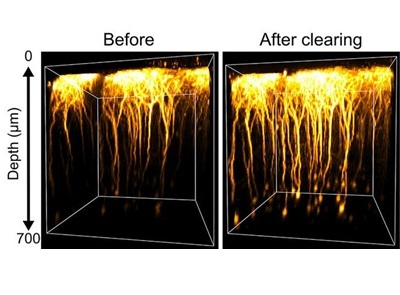
This opens up clear views of layer 5 of the cerebral cortex, where richly branched neurons help reveal how the brain processes information and translates neural activity into action. Before SeeDB-Live, crisp images at this depth were difficult to obtain.
Moreover, as the extracellular fluid washes out SeeDB-Live within hours, the tissue transparency returns to its original state. Because this causes no permanent changes, the same mouse can be imaged repeatedly, safely, to track brain activity over time.
Researchers expect SeeDB-Live to enhance deep fluorescence imaging for understanding brain integrative functions. They say it may also help evaluate 3D tissues and brain organoids for drug discovery research.
“I feel we have not yet fully materialized its potential,” said Inagaki, adding that future efforts will focus on less invasive delivery methods to improve penetration for deeper imaging and better functional analysis of brain activity.