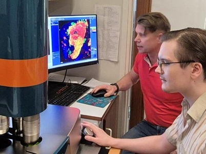
PhD student Joakim Lehrstrand (foreground) and Professor Ulf Ahlgren at a so called light sheet fluorescence microscopy, one of the techniques they use to create three-dimensional images of the pancreas in diabetes. Credit: Björn Morén
Using advanced imaging technologies, researchers at Umeå University have created the first complete 3D map at a microscopic resolution of an entire pancreas from a donor with late‑onset type 1 diabetes. The results show that insulin-producing cells can remain long after the onset of type 1 diabetes—a finding that suggests the disease progression is more complex than previously assumed.
The analysis, published in Science Advances, revealed that while traditional islets of Langerhans were largely depleted of β-cells, a substantial number of insulin‑producing cells still remained outside the islet structures. These were primarily found as individual cells or as small clusters of β-cells, in which the cells were distanced from all other endocrine cell types. In total, the researchers identified hundreds of thousands of insulin‑positive objects.
The researchers argue that traditional islet-focused analyses risk underestimating how many β-cells actually survive in type 1 diabetes. The new findings point to a previously overlooked cellular reservoir that could, in the long term, become a target for novel therapeutic strategies.
“The ability to study individual cells throughout an entire organ and from all angles has the potential to change how we think about β‑cell loss,” said study author Ulf Ahlgren, professor in the Department of Medical and Translational Biology at Umeå University. “If certain regions of the pancreas promote β‑cell survival, understanding these microenvironments could help guide the development of therapies that stabilize, or even expand the remaining β-cells in type 1 diabetes.”
The research team believes that whole‑organ 3D imaging will become a key tool in future studies of type 1 diabetes and other pancreas-related diseases, such as type 2 diabetes and pancreas cancer.
The new method makes it possible to identify specific regions or even individual cells throughout the entire organ, something that has previously been extremely difficult using conventional techniques. These regions can then be isolated for further molecular analyses.
“Hopefully, this will help us understand whether and how β-cells and their microenvironment differ within the pancreas in diabetes,” said Ahlgren.
Data from Umea University