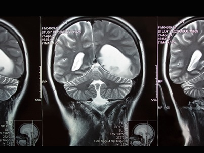
A University at Buffalo-led team has flipped the script on multiple sclerosis, using AI to reveal patient brain lesions doctors knew were present but couldn’t see on scans.
It’s long been known that the gray matter of the brain plays a key role in MS disease progression and cognitive impairment, but because magnetic resonance imaging (MRI) has only been able to detect lesions in white matter, neither clinicians nor researchers have had a way to detect or monitor gray matter (cortical) lesions.
Now, in a paper published in Communications Medicine, researchers have found a way to use artificial intelligence to reveal these otherwise invisible cortical lesions by reviewing existing MRI scans.
The AI approaches the researchers used, building on work from the co-authors from the Netherlands, were designed to extrapolate vital information from the relationships between multiple images that can’t be seen on a single image.
The researchers combined multiple image-processing techniques, including a new one they developed called MMCLE, or multimodal cortical lesion enhancement. They then applied these techniques to MRI scans from the large, phase III FDA regulatory ORATORIO clinical trial, a study of the MS drug Ocrelizumab that included more than 700 participants.
The team found that while individual images of a patient’s brain revealed mostly white matter lesions, once they applied the AI-based image processing methods to multiple different contrast images, they were able to see anywhere from 15 to 20 cortical lesions for each patient—more than 11,000 for the whole dataset.
“If you look on the original scans, you generally can’t see the cortical lesions, but generative AI is very powerful because it can look between the scans and detect tiny differences between them,” said Michael Dwyer, first and corresponding author on the paper, associate professor of neurology and biomedical informatics in the Jacobs School of Medicine. “Because it sees those minor discrepancies, AI can reveal that there’s something going wrong there, that the tissue is not behaving like healthy tissue. The trained models can view multiple MRI images together and synthesize them and what had been missing.”
Data from University at Buffalo