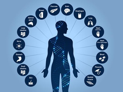
Salk Institute researchers profiled the 3D genome structure of cells from 16 different tissues from the human body to create an atlas that will help scientists study human health and disease. Credit: Amy Cao, Salk Senior Illustrator
Every cell in the human body carries the same three billion letters of DNA, yet a neuron, a heart muscle cell and a pancreatic beta cell each read that code completely differently. Researchers at the Salk Institute have now built the first atlas that maps two of the major systems behind that trick—three-dimensional genome folding and DNA methylation—together in the same single cells across the human body.
The new resource profiles 86,689 cells drawn from 16 human tissues, identifying 35 major cell types and 206 subtypes.
For the study, published in Science, the researchers measured both epigenetic layers simultaneously rather than separately, which allowed them to directly compare what each one reveals about a cell's identity and—importantly—where the two pictures disagree.
Using tissue from the heart, brain, lungs, stomach, skin and other organs, the team catalogued more than 1.36 million differentially methylated regions and 283,606 differential chromatin loops.
When the researchers layered known disease-risk genetic variants onto this map, clear patterns emerged: variants tied to blood-glucose regulation clustered in endocrine cells, atrial fibrillation variants in heart muscle cells, balding-related variants in skin fibroblasts, and bipolar disorder and schizophrenia variants in excitatory and inhibitory neurons.
“A lot of the genetic variation that predisposes someone to disease is in non-coding parts of the genome,” said co-corresponding author Jesse Dixon, MD, PhD, associate professor at Salk. “By adding in the 3D genome aspect, we can potentially bridge that gap—connecting noncoding variations with the genes they affect in specific cells and tissues.”
Cell by cell
The scientists discovered, much to their surprise, that the two epigenetic signals don't always agree.
In skeletal muscle, some fibers looked like fully mature muscle cells based on their 3D genome folding, yet still carried the methylation signature of muscle stem cells. The team says this mismatch likely reflects cells caught mid-transition, with genome architecture changing faster than methylation catches up.
Similar disagreements turned up in Schwann cells and placental trophoblasts, suggesting the two systems update on different timelines as cells change state. The atlas also revises a long-standing assumption about “non-CG methylation,” an unusual form of methylation previously thought to be largely confined to brain cells and stem cells. The study shows that it carries cell-identity information across many human tissues, including muscle, pancreas and immune cell types, at lower but biologically meaningful levels.
In a companion paper in the same issue of Science, Bing Ren from the New York Genome Center and Columbia University used advanced single-cell analysis to examine postmortem hippocampal tissue from 40 neurologically healthy adults between the ages of 20 and 95. Ren and team found that microglia—the brain's resident immune cells—progressively decline between roughly age 50 and 75. As they disappear, they're replaced by cells bearing inflammatory markers and other traits more typical of immune cells found in the bloodstream, rather than the brain.
The findings challenge the long-held assumption that microglia, which form during embryonic development, simply renew themselves throughout a person's life. The researchers also observed that cells maintaining the blood-brain barrier deteriorate with age, and that many brain cell types show a broad, coordinated breakdown in genome architecture over time.
"Gene expression tells us what a cell is doing today, but epigenetic signatures preserve information about where a cell came from," said Nathan Zemke, director of single-cell genomics at the UC San Diego Center for Epigenomics and the study's first author. "By combining these approaches, we uncovered a major shift in the identity and lineage of immune cells in the aging human brain that gene expression data alone would not have revealed."
A public resource
To share the data, the group built a free interactive online browser containing 195 billion methylation measurements and 18 billion chromatin contacts, intended both as a reference for gene-regulation researchers and as training data for AI models that predict how genetic variants affect disease risk. Atlases like this one provide the labeled, cell-type-resolved training data that AI models need to make accurate predictions—a bottleneck that has historically limited the field.
The 4D Nucleome consortium's next goal is to extend this kind of mapping into a true fourth dimension—time—tracking how genome structure and chemistry shift as cells develop, age and respond to disease, with this cross-tissue atlas serving as a reference point for those future studies.