Multispectral imaging systems are used by the food industry, the pharmaceutical industry, and in medical, biology, and chemistry labs for analysis and quality control. Types of multispectral imaging include NIR (near infrared), which can provide a quick and accurate analysis of samples within the 700–2500 nm wavelength range, and Raman, which measures the scattering of near infrared, visible, or near ultraviolet light. In both cases, the photons that are emitted and absorbed by the sample allow identification of specific molecules.
Considerations for purchasing a Multispectral Imaging System
Sensitivity, specificity, and resolution of images can vary between chemical imaging systems. Another feature to be aware of is what type of imaging the system is capable of and whether other types of imaging can be integrated into the system.
Discover and compare Multispectral Imaging Systems:
|  |  |  |
| Company | Oxford Instruments | Oxford Instruments | Oxford Instruments |
| Item | RISE Microscopy – Raman Imaging-SEM | ParticleScout – Raman-based Particle Analysis | TrueSurface – Topographic Raman Imaging |
| Citations | | | |
| Catalog Number | RISE™ Microscopy | ParticleScout™ | TrueSurface™ |
| Price | | | |
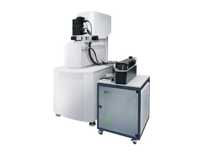
| Description | RISE Microscopy is a novel correlative microscopy technique that combines confocal Raman Imaging and SEM to link ultra-structural surface properties with molecular compound information.
The RISE Microscope combines all features of a stand-alone SEM and the witec360 confocal Raman imaging microscopeRISE Microscopy is a novel correlative microscopy technique that combines confocal Raman Imaging and SEM to link ultra-structural surface properties with molecular compound information.
The RISE Microscope combines all features of a stand-alone SEM and the witec360 confocal Raman imaging microscope within one instrument.
- Quick and convenient switching between Raman and SEM measurement.
-
Automated sample transfer from one measuring position to the other.
-
Integrated software interface for user-friendly measurement control.
-
Correlation of the measurement results and image overlay.
-
No compromise in SEM and Raman imaging capabilities.
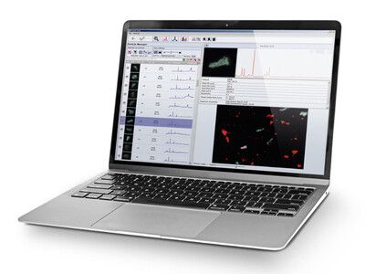
... Read More | ParticleScout is an advanced particle analysis tool for witec360 Raman microscopes that finds, classifies and identifies microparticles over even large sample areas.
User-defined categorization of particles by shape and size offers access to wide-ranging properties, and the results of the ParticleScout is an advanced particle analysis tool for witec360 Raman microscopes that finds, classifies and identifies microparticles over even large sample areas.
User-defined categorization of particles by shape and size offers access to wide-ranging properties, and the results of the measurement are then presented in a report that provides a comprehensive description of the sample.
ParticleScout features the most flexible and useful sample survey capabilities available.
- White-light Microscopy: Bright field/dark field, transmission/reflection.
-
Image Stitching: Scans of many sample areas can be combined into one measurement for a large-area overview of widely distributed particles.
-
Focus Stacking: This unique capability dramatically increases the depth of focus for sharp and defined particle imaging.
-
Integration time optimisation – reduces measurement time and minimizes the effects of fluorescence.
-
Vignetting correction – ensures uniform brightness throughout the image plane.
-
Smart zoom – displays particle information dynamically depending on viewed area.
-
Sample area targeting – allows the selection of multiple regions of interest for each investigation.
-
Dark-field, bright-field, epifluorescence and transmission sample illumination – provides the best option for each type of particle.
-
Round sample wedge sectioning – allows the selection of an area for data acquisition, typically on a filter, that can be extrapolated to represent the whole.
-
Smart separation of particles – differentiates materials in densely packed, heterogeneous samples.
-
• Quantitative report formatting – presents data using table, bar graph histogram and pie chart templates for ease and clarity.
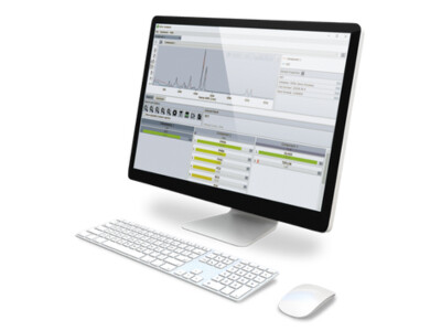
... Read More | The patented TrueSurface Microscopy option for witec360 microscopes enables confocal Raman imaging guided by surface topography to maintain an optimal focus throughout a measurement.
The pioneering topographic Raman imaging technology uses an integrated optical profilometer to provide one-pass The patented TrueSurface Microscopy option for witec360 microscopes enables confocal Raman imaging guided by surface topography to maintain an optimal focus throughout a measurement.
The pioneering topographic Raman imaging technology uses an integrated optical profilometer to provide one-pass simultaneous operation.
- Enables 3D chemical characterisation of rough, inclined or irregularly shaped samples.
-
• Compensates for variations during measurements with long integration times.
-
• Features variable offset from the sample surface.
-
• Integrates seamlessly with Raman, PL, white-light microscopy.
-
• Further reduces sample preparation effort.
... Read More |
| Type | Inquire | Inquire | Confocal Raman Imaging |
| Wavelength Range | UV-VIS-NIR | UV-VIS-NIR | UV-VIS-NIR |
| Scan Time | Inquire | Inquire | 1 to 50 ms |
| Spectral Bandwidth | UV-VIS-NIR | UV-VIS-NIR | UV-VIS-NIR |
| Detector(s) | CCD/EMCCD | CCD/EMCCD | Optical Profilometer |
| Get Quote | | | |