Deep vein thrombosis (DVT) is a common and potentially life-threatening condition that is difficult to diagnose with a typical ultrasound. Treating clots in deep veins can also be challenging, with potential side effects from blood thinning medications or invasive surgical procedures. A team of researchers from the Penn State College of Engineering developing an improved imaging technique for diagnosing DVT made an unexpected discovery while testing their method, which may lead to new treatment options for the condition.
The team previously developed a particle-based imaging approach in 2017, designing nanopeptisomes (NPeps) that would bind to the surface of clots, making them easier to see using an ultrasound. The NPeps consist of a shell around a droplet of fluorine-based oil that turns to gas and appears as a “bubble” under an ultrasound, showing where a clot is forming.
The researchers tested their method on bovine veins, using an enzyme to induce clot formation and applying the NPep particles in order to view the clots. However, the researchers found that they only saw clot formation about 30% of the time, even though the enzyme generally induces clot formation 100% of the time, according to lead researcher Scott Medina. The team continued testing the method and found they would lose the signal of the “bubbles” under the ultrasound after 15 minutes during each test, suggesting that the particles may be breaking down the clots. The study was published in Advance Healthcare Materials.
“We think that once our particles start to decorate the clot, they saturate the surface and inhibit the mechanisms of further clot growth,” said Medina. “And under the ultrasound, the particles are disrupting the clot or inhibiting its mechanism to persist. While we don’t understand the underlying mechanism yet, it’s clear that these particles can image and help treat clots in real time.”
The team plans to further investigate how the particles disrupt the clots and also plan to develop more control over how the particles behave. They hope further development of the technique could lead to new opportunities for non-invasive, image-guided DVT interventions.
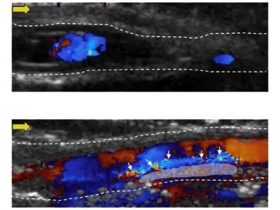
Photo: Penn State researchers developed particles that target blood clots and visualize their structures well compared to traditional ultrasound imaging. These images show blood flowing from left to right: The top image does not have the particles, while the bottom one does. Credit: Penn State/Scott Medina