Chest X-rays and computed tomography (CT) scans are often use to screen patients for lung diseases such as chronic obstructive pulmonary disease (COPD), but fine distinctions between healthy and diseased tissue are challenging to pick up on a conventional X-ray image, and CT exams can be time-intensive, costly and require relatively high amounts of radiation. Researchers from the Technical University of Munich (TUM) have now presented the results of an initial clinical patient study involving a new form of diagnostic X-ray technology that provides better clarity, faster, and at a lower cost and radiation dosage.
Dark-field X-ray imaging utilizes the phenomenon of scattering in a similar manner to dark-field microscopy. Rather than measuring the attenuation of X-rays, as is done in conventional X-ray imaging, dark-field X-ray imaging uses the coherent small-angle scattering of X-rays at microscopic structures, allowing the technology to visualize the fine interfaces between air and tissue. This is particularly helpful for imaging the lungs, as clinicians can clearly distinguish between intact, air-containing alveoli and alveoli that are damaged, according to Franz Pfeifer, director of the Munich Institute of Biomedical Engineering at TUM.
This technique provides better information than a conventional chest X-ray for early detection of diseased alveoli and emphysema, and requires only one exposure, rather than several exposures needed to capture multiple images in a CT exam. The total radiation dose for one exam is estimated to be just 1/50th that of CT, Pfeiffer said. The exam also only takes about 7 seconds to perform.
In the initial clinical patient study, patients with healthy lungs, early signs of emphysema and moderate-to-severe emphysema underwent a conventional chest X-ray, a dark-field chest X-ray and a chest CT scan. This study represented the first time the new technology was used in human patients. The dark-field images provided better structural detail of the lungs for diagnosing the symptoms of COPD, and the information provided by the images were found to be consistent with the findings from the CT exams. The results were published in The Lancet Digital Health.
“Given the close connection between the alveolar structure and the functional condition of the lung, this ability is of great significance for pulmonary medicine,” said Dr. Alexander Fingerle, a senior physician at the Department of Diagnostic and Interventional Radiology at TUM’s university hospital and a coauthor of the study. “In the future dark-field X-rays could help improve early detection of COPD and other respiratory ailments.”
The researchers are interested in conducting further research to investigate the ability for dark-field X-ray imaging to be applied to other lung conditions such as pulmonary fibrosis, pneumothorax, lung cancer, pneumonia and COVID-19.
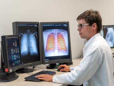
Photo: Co-author Dr. Andreas Sauter evaluating X-ray images in the Institute for Diagnostic and Interventional Radiology in TUM's university hospital Klinikum rechts der Isar. Credit: Andreas Heddergott/TUM