The phenotypes of various key crop plants such as wheat, corn and grapes are both visible to the naked eye and driven by microscopic processes at the cellular and molecular level. Tying together detailed information from plant microstructures and larger scale organs and tissues can be challenging with conventional microscopy methods that require smaller samples and only capture a portion of the larger structures without full spatial context. New 3D X-ray microscopy (XRM) methodologies recently published by researchers at the Donald Danforth Plant Science Center aim to provide scientists a way to capture cellular-resolution, multiscale images of plants with improved depth and range.
The 3D XRM technology allows developmental microstructures, such as meristem cells, to be related to visible traits, such as leaves and flowers, as plants mature. Scanning a plant sample with this technology generates a 3D cell-level map, providing adequate resolution, spatial reference and the ability to identify subcellular regions of interest for further examination through methods such as scanning electron microscopy (SEM). The researchers designed specific methodologies and workflows to obtain the clearest XRM images of specific plant structures for a range of different species, including corn, foxtail millet, soybean and grape.
In addition to imagining developmental structures, flowers, leaves and other “above-ground” organs, the 3D XRM technology can also be used to examine “below-ground” structures such as roots, as well as the fungi and microbes that plants interact with under the soil. This capability can also help researchers to understand and encourage natural production of phosphorus and nitrogen and reduce reliance on chemical fertilizers. This work was published in the journal Plant Physiology.
“The idea of the paper was to give plant scientists working on a variety of relevant plant tissues and species the access to these methods,” said Christopher Topp, who led the research team. “We want to broadly apply 3D XRM to plant systems above and below ground.”
Advanced 3D XRM methods can help scientists better understand how microscopic structures and processes contribute to important macroscale plant phenotypes such as size and color, and how natural phosphorus and nitrogen production can be promoted in a more sustainable way. The researchers are also interested in developing technology to image the 3D structures of fungal networks within soil, and using machine learning to identify structures such as roots and spores from 3D images.
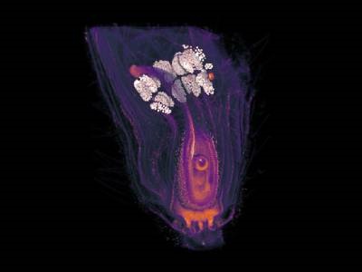
Photo: X-ray microscope 3D volume rendering of a developing soybean flower. Credit: Donald Danforth Plant Science Center