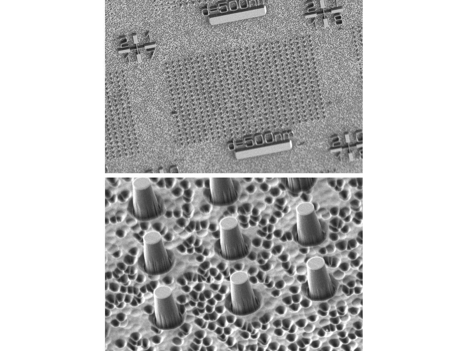
The experimental set-up: A 30-micron-thick diamond membrane with one sensor, on average, at the top of each column, magnified 2,640 times (top) and 32,650 times (bottom). Credit: Weizmann Institute of Science
The ability to locate and map the spins of single electrons could enable a range of applications such as magnetic resonance imaging (MRI) on the nanoscale and characterization of quantum materials. However, determining and visualizing the positions of individual electrons is no easy task, as these subatomic particles are far smaller than what can be seen with even the most advanced microscopes – so small that their exact size can’t even be measured. Despite these challenges, researchers at the Weizmann Institute of Science have now presented a method capable of detecting and mapping individual electron spins, using magnetic fields and an atom-sized quantum sensor.
The method is based on magnetic tomography, and involves the modulation of rotating magnetic fields in the vicinity of a nitrogen-vacancy center, which is an atom-sized defect in a synthetic diamond. The nitrogen-vacancy center serves as a quantum sensor that is highly sensitive to nearby changes – sensitive enough to detect the presence of a single electron. By using changes in the magnetic field vector to manipulate the dipolar coupling reactions between the spin sensor and a nearby electron spin within the diamond, the researchers can determine the position of the electron with high precision. For their experiment, the team used a 30-micron-thick diamond membrane engineered to contain hundreds of nitrogen-vacancy centers within a nanoscale area.
The researchers found that their magnetic tomography method enabled them to determine the location of electron spins with an uncertainty of 0.9 Å, and that electron locations could be determined with nanometer precision up to 10 nm away from the quantum spin sensor. While the technology is still in its early stages, the ability to pinpoint the locations of individual electrons has potential implications for a wide range of applications, as maps of electrons could enable ultra-precise distance measurements when studying the structures of individual molecules, the authors wrote. For example, in pharmaceutical development, the technique could improve the engineering of novel drug molecules. This capability could also expand the application of MRI technology to the nanoscale and improve methods for characterizing quantum networks and materials. This study was published in Physical Review Applied.
“This new method could be harnessed to provide a complementary point of view to existing methods, in an effort to better understand the holy molecular trinity of structure, function and dynamics,” said study co-author Amit Finkler.