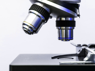
MIT researchers have developed an alternative imaging technique that avoids the need for expensive super-resolution microscopes. By expanding tissue before imaging it, the researchers achieved nanoscale resolution with a conventional light microscope.
In their latest research, the team expanded tissue 20-fold in a single step. "This democratizes imaging," said Laura Kiessling, Professor of Chemistry at MIT.
"Without this method, if you want to see things with a high resolution, you have to use very expensive microscopes. What this new technique allows you to do is see things that you couldn't normally see with standard microscopes. It drives down the cost of imaging because you can see nanoscale things without the need for a specialized facility."
"Twenty-fold expansion gets you into the realm that biological molecules operate in. The building blocks of life are nanoscale things: biomolecules, genes, and gene products," added Edward Boyden, Professor in Neurotechnology at MIT.
The team initially invented expansion microscopy in 2015, a technique that embeds tissue into an absorbent polymer that swells with the addition of water. This swelling separates the biomolecules, allowing for nanoscale imaging with typical lab microscopes.
While the original method achieved a four-fold expansion, the recent technique, published in Nature Methods, achieved a 20-fold expansion in a single step. Once the N,N-dimethylacrylamide (DMAA) and sodium acrylate gel are formed, select protein bonds are broken and water is added to expand the gel.
"This approach may require more sample preparation compared to other super-resolution techniques, but it's much simpler when it comes to the actual imaging process, especially for 3D imaging," said Tay Won Shin, an MIT graduate student.
Using the technique researchers achieved a resolution of around 20 nanometers using a conventional light microscope, allowing structures such as microtubules and mitochondria to be imaged.
"Our hope is that with this new technology, any conventional biology lab can use this protocol with their existing microscopes, allowing them to approach resolution that can only be achieved with very specialized and costly state-of-the-art microscopes," added MIT graduate student Shiwei Wang.