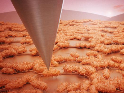
This artistic rendering depicts an atomic force microscopy tip scanning a structured network of bacterial cells with flagella in a honeycomb pattern. High-resolution surface characterization spans a wide field of view. Credit: Adam Malin/ORNL, U.S. Dept. of Energy
DOE scientists at the Oak Ridge National Laboratory have developed a groundbreaking new technique which transforms the capabilities of atomic force microscopy (AFM) from imaging nanoscale featured to capturing large-scale biological architecture. Traditionally, AFM has been limited by a narrow field of view, making it challenging to determine how individual featured wit within larger organizational structures.
By developing a large-area automated AFM platform, researchers were able to overcome this narrow field of view. Published in the journal npj Biofilms and Microbiomes, the new AFM platform allowed the team to connect detailed observations of bacterial biofilms with broader views across a larger area.
“In biofilm research, we’ve often been able to see the trees, but not the forest,” said Ruben Millan-Solsona, a postdoctoral researcher in ORNL’s Functional Atomic Force Microscopy group. “Using the AFM, we could examine individual bacterial cells in detail but not how they organize and interact as communities.”
“This new platform changes that. Now, we can visualize both the intricate structures of single cells and the larger patterns across entire biofilms,” added Liam Collins, an R&D researcher in ORNL's Functional Atomic Force Microscopy group.
Key to the development was the integration of machine learning with the imaging process. “The large-area AFM provides researchers with large-scale, high-resolution views of biofilms,” added Sita Sirisha Madugula, a postdoctoral researcher in ORNL’s Data Nanoanalytics group. “The integration of machine learning allows us to extract meaningful quantitative data from these massive datasets.”
Using the technique the team automatically analyzed over 19,000 cells to generate a detailed map across an extensive surface area, revealing a honeycomb-like pattern of bacteria which are interconnected by flagella.
“Though the biological role of these patterns is still under investigation, they likely play a part in strengthening biofilm cohesion and adaptability,” Collins added.
The AFM method developed marks a significant step forward in the science of biofilm analysis.