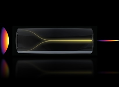
Under the right conditions, a chaotic mess of laser light can spontaneously self-organize into a highly focused “pencil beam.” This schematic shows the pencil beam formation mechanism. Credit: Courtesy of the researchers/MIT
MIT researchers discovered a phenomenon in optical physics that could enable a new bioimaging method that’s faster and higher resolution than existing technology.
Under the right conditions, laser light can spontaneously self-organize into a highly focused “pencil beam.” Using this self-organized pencil beam, the researchers captured 3D images of the human blood-brain barrier 25 times faster than the gold-standard method, while maintaining comparable resolution.
The discovery began with an observation that initially puzzled the researchers. They previously developed a precise fiber shaper, a device that enables them to carefully tune the laser light shining through a multimode optical fiber. Typically, the more power one pumps into the laser, the more disordered and scattered the beam of light becomes due to imperfections in the fiber. But, as power was increased almost to the point where it would burn the fiber, the light did the opposite of what was expected: it collapsed into a single, needle-sharp beam.
To replicate this, the researchers found they had to satisfy two simple, but precise conditions. First, the laser must enter the fiber at a perfect, zero-degree angle. Second, the power must be dialed up until the light begins to interact with the glass of the fiber itself.
When the researchers performed characterization experiments of this pencil beam, it was more stable and high-resolution than many similar beams. They then employed it for biomedical imaging of the human blood-brain barrier. Using the technique, the researchers created an ultrafast, high-precision pencil beam that enabled them to dynamically track how cells absorb proteins in real-time. They captured cellular-level 3D images that were higher quality than with other methods, and generated these images about 25 times faster.
By showing individual cells absorbing drugs in real-time, this technology could help scientists test whether new drugs for neurodegenerative disease like Alzheimer’s or ALS reach their targets in the brain, with greater speed and resolution.
“The pharmaceutical industry is especially interested in using human-based models to screen for drugs that effectively cross the barrier, as animal models often fail to predict what happens in humans,” said Roger Kamm, professor of biological and mechanical engineering at MIT. “That this new method doesn’t require the cells to have a fluorescent tag is a game-changer. For the first time, we can now visualize the time-dependent entry of drugs into the brain and even identify the rate at which specific cell types internalize the drug.”
Data from MIT