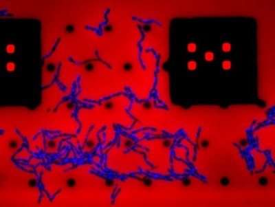
B. subtilis growing under the microscope on a microfluidic chip. The cells are losing their characteristic rod shape because they are being treated with a drug that blocks teichoic acid synthesis. Credit: Felix Barber, NYU/OSU
The rod shape of bacteria is so recognizable that there are even stuffed animals designed after their shape. Now, researchers have discovered what is responsible for that characteristic shape—acids on the surface of the bacteria keep an enzyme at bay that would otherwise turn the cylindrical cells into shape-shifting blobs.
The shape of bacteria is important medically since the cell wall determines cell shape and is the target of frontline antibiotics. A bacterium’s shape dictates how it grows, how it divides, and how it interacts with its environment.
For example, Bacillus subtilis is a rod-shaped bacterium naturally found in soil and in the gut. Considered a probiotic, B. subtilis is used to manufacture a range of foods, antibiotics, skincare and agricultural products. The cell wall of B. subtilis and other Gram-positive bacteria is made up of two components: peptidoglycan, a layer of sugars and amino acids that is primarily synthesized by clusters of proteins called Rod complexes, and long polymeric molecules called wall teichoic acids.
In this study, published in Nature Microbiology, researchers found that eliminating wall teichoic acids in B. subtilis rapidly arrested Rod complexes and simultaneously unleashed the activity of an enzyme called PBP1, which usually plays a minor role in cell wall synthesis by fixing mistakes made by the Rod complexes. This explained why cells turned into blobs—since Rod complexes reinforce the cell wall along its circumference, essentially girdling the cell into a rod shape, whereas PBP1 synthesizes peptidoglycan in random directions, leading to a blob shape.
The next question was how teichoic acids controlled the proteins. Since PBP1 was thought to mend holes in the cell wall, the scientists wondered if it was taking over from Rod complexes when teichoic acid depletion exposed pores in the cell wall. To test this, they developed a new method to measure the cell wall’s porosity with nanoscopic resolution. The team found that nanometer-sized holes appeared within minutes of teichoic acid depletion.
Amorphous growth was not only driven by PBP1—it also required the enzyme. In bacteria lacking it, depleting teichoic acids led to cell wall thinning, an increase in cell wall pores, and a complete arrest of cell growth—all while retaining the bacterium’s rod shape. In addition to PBP1, the researchers found that cell growth without teichoic acids also required a second enzyme, LytE, that chops up the cell wall and is needed for rod-shaped growth.
The findings have implications for other bacteria. For example, Listeria monocytogenes, another rod-shaped Gram-positive bacteria and common culprit in foodborne illness, also loses its shape when teichoic acids are depleted. Studies also show that blocking the synthesis of teichoic acids in methicillin-resistant Staphylococcus aureus (MRSA)—the most notorious antibiotic-resistant bacterium—using an FDA-approved antiplatelet drug can re-sensitize it to antibiotics.
Data from NYU