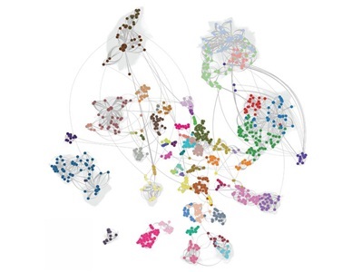
The BICAN consensus basal ganglia cell type taxonomy generated through iterative clustering and cross-species integration of single-nucleus transcriptomic data from human, macaque, and marmoset. Credit: Allen Institute
A global consortium of hundreds of scientists has produced the most detailed cellular atlas of basal ganglia, profiling 25.5 million cells from humans, macaques and marmosets, with mouse data added to the reference.
Basal ganglia, a set of interconnected structures deep in the brain that help control movement, habit formation, decision-making and emotion, has genetic and molecular ties to Parkinson’s disease, Huntington’s disease, schizophrenia, bipolar disorder and addiction.
The atlas is the first major output of the NIH’s BRAIN Initiative Cell Atlas Network (BICAN), which aims to build a complete “parts list” of the human brain. For the studies, published in Cell and other Cell Press journals, researchers from more than 50 institutions across nine countries profiled 21 million human cells, 3 million macaque cells and 1.5 million marmoset cells.
New cell types and disease links
What sets the package apart, researchers say, is that every study used the same cellular framework.
“This community worked together to develop a common description of cell populations, and then everybody used it,” said Ed Lein, executive vice president at the Allen Institute. “We hope this will be a framework for the entire field.”
Allen Institute researchers identified a previously unknown neuron type, STR D2 SMYD2-HTR7, that expresses the serotonin receptor HTR7 and carries elevated risk for schizophrenia and bipolar disorder.
Other studies in the package found key genetic changes in specific basal ganglia cell types that could play a role in autism, and widespread, cell-type-specific changes in gene expression with aging—enough that a person’s age could be predicted from expression patterns across multiple cell types.
In a separate study, scientists developed a “smarter” version of an existing gene therapy that reduced Parkinson’s-like tremors in animal models and was both more effective and less invasive than current approaches.
Following cells from before birth
A UCLA-led team analyzed more than 155,000 nuclei from nine developing human brains, spanning mid-pregnancy through seven months after birth. Combined with earlier developmental and adult data, the analysis covered nearly 287,000 cells.
Using a technology that reads both DNA methylation and 3D genome organization in individual nuclei, the researchers found that cells arising from different parts of the temporary prenatal structures that feed the basal ganglia matured on markedly different schedules—some relatively mature by mid-pregnancy, others still producing immature neurons in infancy.
Spiny projection neurons, a major basal ganglia cell type, matured in an unusually coordinated way, and some showed signs of neuronal activity as early as mid-pregnancy. After birth, oligodendrocytes, the cells that produce myelin, temporarily shifted their DNA into a different 3D configuration linked to greater gene activity as they matured. Spiny projection neurons also showed particularly strong enrichment for genetic variants associated with schizophrenia, with some subtypes tied to bipolar disorder.
“Genetic studies can tell us that particular regions of the genome are associated with disease, but they often can’t tell us which cell type is affected or when during development that genetic risk becomes relevant,” said Chongyuan Luo, associate professor of human genetics at UCLA. “This atlas gives us that cellular and developmental context.” The tissue came from typically developing brains, so the work doesn’t show that these cells cause any disorder.
Which genes stay silent
Two human studies from the New York Genome Center examined both active and repressed genes across more than 1.3 million nuclei. One analyzed 608,000 nuclei from eight basal ganglia regions and identified active and repressive chromatin states across 61 cell groups. Distinct combinations of homeobox transcription factors tracked with neuronal populations, supporting a model in which a homeobox “code” helps define neuronal identity.
The other, covering 750,000 nuclei from 18 brain regions, found that repressive chromatin preserves traces of developmental history: glial cells kept signatures of their forebrain or hindbrain origins, and a class of very long-range chromatin interactions specific to mature neurons was tied to Polycomb-mediated repression.
The team also built EpiBRAIN, a deep-learning model that predicts how noncoding genetic variants affect gene regulation in specific cell types. In benchmarking, it outperformed the Borzoi and AlphaGenome models at identifying variants likely to influence gene expression in the basal ganglia, and variants it prioritized accounted for a larger estimated share of schizophrenia heritability than those chosen by Borzoi. Companion studies mapped more than 100 cell types across roughly 81% of the mouse genome using 2.5 million nuclei, and built a standardized multispecies reference spanning humans, macaques, marmosets and mice.
Mapping where cells sit
At UC Irvine, researchers combined two spatial transcriptomics technologies, MERFISH+ and Stereo-seq, to map about 8 million cells from four neurotypical donors, identifying 60 cell types and showing how they form cellular communities. Different cell types occupied distinct areas of the basal ganglia. For example, some supporting cells clustered along compartment borders, while certain neurons more abundant in primates appeared only within particular compartments.
What comes next
The researchers stress that the atlases are reference resources and that their clinical value depends on follow-up experiments to test how specific variants affect cell function.
The next goal is the rest of the brain.
“It’s the sum output that really differentiates this set of studies,” said Lein. “All of them largely agree with one another. This molecular classification works across different data types and labs. That really gives us confidence that this gets to the ground truth of the organization of the brain.”