Featured Article
Keeping cells and tissues in place is a must for microscopy
Watching a histologist mounting tissues on slides quickly reveals the complexity of the task. All goes for naught, though, if the sample won’t stay on the slide. “It is not common for scientists to run into problems with keeping cells on microscope slides for simple staining processes, such as hematoxylin and eosin—H&E—staining procedures,” says Lise Duran, vice president R&D at Leica Biosystems (Richmond, Ill.). Some procedures, though, make cell adhesion very complicated. “Formerly,” says Kimberly A. Glover, life sciences product manager at Polysciences (Warrington, Penn.), “there was a concern with cells and tissue coming off the slide when preforming ‘harsh’ procedures, such as in situ hybridization techniques and HIER [heat-induced epitope retrieval] for immunohistochemistry.”
Problems arise from other conditions, such as high pH, elevated temperature and agitation. Duran says, “Immunohistochemistry techniques as well as many special stains, such as silver, will likely cause certain cells to come off slides.”
Some tissues are more difficult to adhere to slides than others. Duran points out that fatty tissues, including brain and breast samples, tend to fall off of slides. “These tissues do not typically adhere to glass,” Duran says. “Also, any calcified tissue or tissue with excess amounts of keratin, such as bone, are hard and do not lay flat on the glass surface.” Duran adds, “As an example, approximately 50% of toenail samples will fall off glass slides during staining.”
A variety of new products offer solutions to keep cells or tissues on microscope slides.
Opposites attract
The ionic distribution across cell membranes gives them a charge, and histologists can take advantage of this. “Since most cells are negatively charged, microscope slides can be treated with positively charged compounds,” Duran explains. “These compounds adhere well to the glass surface and can ionically bond to the membrane surface of the cell, and once bound, the cells are more firmly attached.”
Suppliers give microscope slides a positive charge by coating them. Some of the most common coatings include functionalized silanes, poly-L-lysine and polymers functionalized with quaternary ammonium groups. Mathew Griffin, technical product specialist at Polysciences, adds, “The addition of more amines, such as by tethering polyethylenimine, PEI, could strengthen the bond.”
Several vendors make silane-treated slides for microscopy. Polysciences offers Tissue Tack slides treated with a specially formulated aminoalkylsilane. These slides provide a positively charged surface, which permits instant coupling of negatively charged tissue sections. Glover says, “The resultant bond stands up to the very aggressive solutions used in in situ hybridization procedures.” StatLab (McKinney, Tex.) HistoBond adhesion slides work well for “routine histology, frozen sections, special stains, as well as [immunohistochemistry],” according to the company website. These silane-treated slides come in eight colors, which can be used to code for cell type, treatment and so on. The colored section is also made to work well with Leica or Sakura (Torrance, Calif.) automated slide printers.
HistoBond adhesive microscope slides from Paul Marienfeld GmbH (Lauda-Königshofen, Germany) also include a silane-treated surface, which it promotes for in situ hybridization techniques. The company website notes that, “even problematic tissues such as brain or spinal cord can be processed with the aid of HistoBond slides in routine histology.” Light Labs (Dallas, Tex.) uses a silane coating on its StarFrost adhesive slides. The company’s website notes that the silane only goes on one side of the slides, so they don’t stick where you don’t want them, like to the benchtop.
Tekdon (Myakka City, Fla.) coats its adhesive slides with amino groups. Company literature states that this ensures “a firm electrostatic attraction to the slide of frozen tissue sections, cell and cytology preparations binding them to the glass surface.” In particular, the company claims that this keeps tissue sections and cells from “floating off the slide during frequent washing when carrying out immune-histochemical staining.”
Several other companies also use amino groups to make slides more adhesive. These include Polysine microscope adhesive slides from Thermo Fisher Scientific (Waltham, Mass.), Poly-L-Lysine coated microscope slides from Polysciences and Poly-Prep slides from Sigma-Aldrich (St. Louis, Mo.).
Most vendors suggest that their adhesive slides work well for various procedures. IHC World (Woodstock, Md.) markets its TruBond microscope slides for manual and automated immunohistochemistry, immunocytochemistry and immunofluorescence.
The details matter
The complexity that can arise from binding cells to slides can be easily seen in a discussion started by Harrie Verhoeven of Wageningen University on ResearchGate.net on February 4, 2014. He wrote, “We are working with single cell layers on microscope slides. The cells need a surface coating to adhere to the glass, which is currently a polylysine 140-kD variant.” Despite adding that he used new and extensively cleaned slides, he still ran into problems with attachment. When he asked for help, scientists from around the world replied.
Louise Elizabeth Smith of the University of South Australia pointed out that it depends on the cell type, but, she adds, “As a general rule, if the biological methods aren’t working and you have access to a physics or engineering department with a plasma reactor, a short oxygen or air plasma should improve adhesion.” She said that will make the surface more hydrophilic and gives the surface “a small amount of topography to increase adhesion.” If that doesn’t work, she suggested using a coating like collagen.
Getting the right bond can require a balance. As Smith wrote, “If your glass is too hydrophobic or too hydrophilic, you will struggle to get any biomolecule to adsorb.”
Maryline Christine Bossus from the University of Arkansas suggested, “If you add CaCl2 (final concentration around 3 mM) and MgCl2 (at about 11 mM) in the cell solution, that will help the cell layer to adhere to the slide.” Not every idea works, though, and Verhoeven replied that his cells could not be exposed to high concentrations of CaCl2.
Others recommended solutions that typically appear in some cell-culture media. Virgilio J.J. Cadete of the University of Montreal pointed out that adding fetal bovine serum to the media improved the adhesion of his cells.
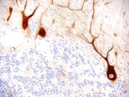
 Immunohistochemistry on human cerebellar tissue mounted on Leica X-tra slides reveals the neurofilaments. (Image courtesy of Leica Biosystems.)
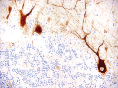
Some of the experts even provided quite detailed suggestions. Ali Moraid of Malaysia’s University of Malaya suggested this pretreatment: Put them on 1 molar hydrochloric acid at 60° Celsius in a magnetic stirrer overnight, then 6 hours in the stirrer in double distilled water, then 6 hours in 70% ethanol followed by drying them in a laminar-flow hood. That’s just to get the slides clean and sterile. Then, Moraid coats the slides with a concoction that includes collagen and fetal bovine serum.
Some of the suggestions got even more specific, such as basing the slide coatings on what genes the cells express. That’s pretty pointed advice to make cells stick!
Teaming up on adhesion
Making the most effective microscope slides takes a team. Duran explains, “We have subject matter experts [who] can evaluate the effective adhesion of cells to glass surfaces.” Duran adds, “These include cell biologists, histologists, material scientists and engineers who routinely monitor the quality of our slides and stay abreast of new technologies to improve cell adhesion.”
It can take a team of supplier experts plus scientists interacting around the world to get the best adhesion of specific cells to microscope slides for particular applications, but the results can make up for all of the effort.
Mike May is a freelance writer and editor living in Ohio. He can be reached at [email protected].