Acquiring detailed 3D image information from a microscope typically involves the data-intensive process of assembling hundreds of 2D images into a stack, which is then processed and rendered by a computer. While 3D reconstructions of microscope images are a valuable resource for researchers, this reconstruction method through focus stacking can be costly and time-consuming. A team of researchers at UT Southwestern recently developed a new, cost-effective method that can enable real-time 3D observation of specimens through modification of common fluorescence microscopes.
The UT Southwestern scientists discovered the method while de-skewing a 3D image stack produced from two light-sheet microscopes. While experimenting with an optical de-skewing method they noticed that de-skewing the stack incorrectly caused the projected image to appear to rotate. The scientists then developed this discovery into a system that allows for real-time multiangle projection imaging. This method involves rapidly imaging a sample from two perspectives and placing two rotating mirrors in front of the camera of the microscope system. The result is a system that allows for interactive visualization of the sample in three dimensions.
The team managed to reproduce this projection imaging system using light-sheet, oblique plane and spinning disk confocal microscopes, and tested it by imaging specimens including cancer cells, neurons and zebrafish embryo. By rapidly imaging the samples, the scientists could observe calcium signaling between neurons and the beating of the zebrafish’s heart. The research was published in Nature Methods.
“We can do all this in real time, without any noticeable delay. Surprisingly, we can look from different angles ‘live’ at our samples without rotating the samples or the microscope,” said corresponding author Reto Fiolka. “We believe this invention may represent a new paradigm for acquiring 3D information via a fluorescence microscope.”
The researchers note that their method both reduces data overhead and accelerates 3D imaging by a factor of 100 or more, which opens up avenues for advanced microscopy in a range of life science research areas.
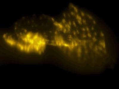
Photo: Microscopy image of a zebrafish embryo. Credit: UT Southwestern Medical Center