The ability to map and measure the vasculature of the brain down to the smallest capillaries can help improve prevention, detection and treatment of cardiovascular disease, aneurysms and stroke. Fluorescent dyes used to map blood flow have the disadvantages of being toxic and potentially affecting blood vessels, distorting results, while genetically modified animal models designed to be imaged without dyes can be an expensive alternative. A team of researchers from the Skolkovo Institute of Science and Technology (Skoltech) and Saratov State University have designed a less costly alternative to visualizing blood flow with fluorescent dyes, utilizing dye-less optical microscopy and strategic image processing to track single blood cells moving through capillaries.
The new method involves a frame-by-frame threshold filtering process, in which each image within a series captured by an optical microscope is processed using a technique called adaptive Niblack filtration. By filtering every frame in this manner, the researchers were able to visualize not only the movement of blood through capillaries, but differentiate single red blood cells in the images. Using this detailed series of images, the researchers could accurately assess the blood flow rates within vessels using particle image velocimetry, according to lead author Maxim Kurochkin, of Skoltech.
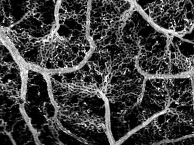
The researchers demonstrated their method by mapping the vascular networks of a mouse brain and chicken embryo, showing how blood flow within the tiniest capillaries of the models could be visualized. Additionally, the researchers showed the method could be used in a more complex model by mapping the vasculature of a rat brain. The research was published in The European Physical Journal Plus.
The team’s method can directly provide information about blood flow rate and vessel diameter, “but once you have that, you can try and extract more information: vessel elasticity, membrane stiffness, blood pressure and viscosity,” Kurochkin explained. “Physiologists building on our work can use these parameters to create blood circulation models, testable against experimental measurements from pressure and viscosity sensors, for example.”
Detailed vasculature models can not only help predict how blood flow is affected by vessel dilation, constriction or obstruction caused by cardiovascular disease, but also aid in a better understanding of blood vessels developed by tumors, how infectious disease affect blood flow and how new drugs may travel through the bloodstream.
Photo: A reconstructed map of the blood vessel network in a chicken embryo obtained by adaptive frame-by-frame threshold filtering of a series of images of moving red blood cells. Credit: Maxim Kurochkin/Skoltech