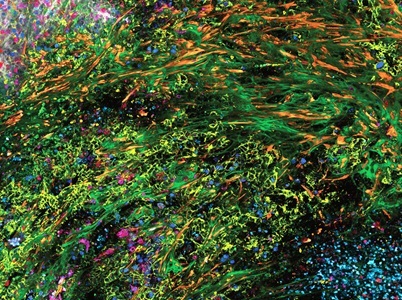
3D high-plex imaging in Cancer Immunology. Overview of a pancreatic tumor section (1.8 x 0.8 mm) in mouse model, labeled with 15 markers and imaged in one pass using STELLARIS with SpectraPlex. Credit: Kunz L., Speziale D., et al., Nat. Methods (2024). https://www.nature.com/articles/d42473-024-00260-7
In recent years, ‘omics techniques have taken a central role in life science research. Understanding the spatiotemporal relationships between various biological components has become crucial for elucidating biological functions in both healthy and diseased states.
To address this, Leica Microsystems has released SpectraPlex, a 3D high-multiplex solution for spatial discoveries on the STELLARIS confocal platform. With a fully integrated workflow, SpectraPlex enables scientists to capture detailed information at the right resolution and in 3D, with 15+ markers in one go, significantly surpassing conventional multi-color imaging. In addition, SpectraPlex offers an offline option to design experiments and explore optimal dye combinations to create panels for high-multiplex imaging. Defining a panel triggers real-time calculations, forming the basis for suggested microscope settings to maximize the signal-to-noise ratio and minimize crosstalk. Experienced users can still finetune these settings to accommodate sample-specific variances.
In SpectraPlex, the unmixed data along with the corresponding raw images is automatically generated to facilitate further analysis and interpretation. Users will benefit from tailored segmentation and downstream analysis for accurate interpretation of high-resolution, 3D data for over 15 labels, all powered by Aivia, Leica Microsystem’s cutting-edge AI image analysis software.