3D-bioprinting is an advanced manufacturing technique capable of producing unique body tissue models using inks embedded with living cells. Models printed using 3D bioink are designed to closely mirror the structure and composition of their natural counterparts; however, realistic 3D-bioprinted models are difficult to produce because the printing process can damage living cells, and higher densities of embedded cells are difficult to print into complex microstructures. Biomaterials engineers at Texas A&M University have recently developed a more biocompatible, structurally stable and printable bioink and leveraged its unique properties to print a realistic blood vessel model with a high density of native endothelial and vascular smooth muscle cells.
The researchers used a combination of polymers and 2D nanosilicates to create a highly-viscous, colloidal bioink material they called nanoengineered extracellular matrix (nECM) to achieve their desired mechanical properties while allowing for cellular interaction and protecting the embedded cells from damage. The 2D nanosilicates interact with the polymer components to provide both reinforcement for the cells during the printing process and help retain the shape of the ink in order to form complex, realistic vascular structures. The team was able to print a 3D vascular model using their new ink and found that the model could accurately mimic thromboinflammation in response to cytokine stimulation. The study was published in Advanced Healthcare Materials.
“A remarkably unique characteristic of this nanoengineered bioink is that regardless of cell density, it demonstrates a high printability and ability to protect encapsulated cells against high shear forces in the bioprinting process,” said corresponding author Akhilesh Gaharwar. “Remarkably, 3D-bioprinted cells maintain a healthy phenotype and remain viable for nearly one month postfabrication.”
3D-bioprinted blood vessel models made from advanced bioinks like nECM can aid in the study of vascular diseases like aneurysms, peripheral artery disease and blood clots, which are leading causes of death worldwide. The availability of an accurate tissue model allows for better precision in developing drugs in vitro with results that are reproducible in vivo, and the ability to produce models using a printer can increase availability of these valuable tools.
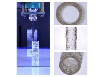
Photo: These images depict the fabrication of a 3D-bioprinted vascular model with native endothelial and vascular smooth muscle cells. Credit: Dr. Akhilesh Gaharwar/Texas A&M Engineering