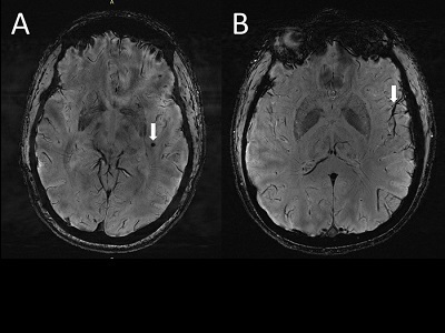
(A) Cerebral microbleeds (CMB) visualized as round, dark lesions (arrow) on SWI sequence in the left temporal lobe in a migraine case with aura. (B) Asymmetry in the appearance of the cortical vessels is more prominent on the left side (arrow) ipsilateral to the CMB. Credit: RSNA and Wilson Xu
For the first time, a new study has identified enlarged perivascular spaces in the brains of migraine sufferers.
Perivascular spaces are fluid-filled spaces surrounding blood vessels in the brain. They are most commonly located in the basal ganglia and white matter of the cerebrum, and along the optic tract. Perivascular spaces are affected by several factors, including abnormalities at the blood-brain barrier and inflammation.
“In people with chronic migraine and episodic migraine without aura, there are significant changes in the perivascular spaces of a brain region called the centrum semiovale,” said study co-author Wilson Xu, an M.D. candidate at the University of Southern California in Los Angeles. “These changes have never been reported before.”
For the study, which will be presented next week at the annual meeting of the Radiological Society of North America (RSNA), participants included 10 people with chronic migraine, 10 with episodic migraine without aura, and 5 age-matched healthy controls.
Statistical analysis revealed that the number of enlarged perivascular spaces in the centrum semiovale (central area of white matter) was significantly higher in patients with migraine compared with healthy controls. In addition, enlarged perivascular space quantity in the centrum semiovale correlated with deep white matter hyperintensity severity in migraine patients.
The researchers hypothesize that significant differences seen might be suggestive of glymphatic disruption within the brain. The glymphatic system is a waste clearance system that utilizes perivascular channels to help eliminate soluble proteins and metabolites from the central nervous system.
However, whether such changes affect migraine development or result from migraine is unknown. Continued study with larger case populations and longitudinal follow-up will better establish the relationship between structural changes and migraine development and type.
“The results of our study could help inspire future, larger-scale studies to continue investigating how changes in the brain’s microscopic vessels and blood supply contribute to different migraine types,” Xu said. “Eventually, this could help us develop new, personalized ways to diagnose and treat migraine.”