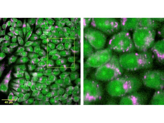
Researchers developed a Raman microscope that can acquire information hundreds of times faster than a conventional Raman microscope. This extra speed makes it possible to acquire large-area hyperspectral images of living cells, such as the ones seen here. Credit: Katsumasa Fujita, Osaka University
Raman microscopy enables high-resolution spectral images to be obtained, combining visual and spatial information with chemical information in a range of applications including life science. However, the application of Raman microscopy in clinical diagnostics has been held back due to the lengthy imaging speeds required to achieve excellent spectral resolution. A team including researchers from Osaka University and the Kyoto Prefectural University of Medicine have now demonstrated new Raman spectral imaging technology that speeds up the process by hundreds of times, enabling high-throughput, high-resolution chemical imaging of large sample areas that could be applied to clinical diagnostics.
The new Raman microscopy approach, known as multiline illumination confocal Raman microscopy, involves the use of a cylindrical lens array to generate multiple line-shaped laser beams from a single expanded beam. This enables the simultaneous irradiation of about 20,000 points within a sample, and the Raman scattering spectra generated from these positions are recorded in a single exposure. A two-dimensional hyperspectral Raman image can be reconstructed by scanning the beams across the sample. Key to the success of the method were the use of a custom spectrophotometer equipped with an optimized array of confocal slits to simultaneously acquire 20,000 spectra, optical filters to prevent spectral overlap and and a high-sensitivity, low-noise CCD camera with a large number of pixels.
The researchers tested their microscope’s ability to acquire measurements and images of live cells and tissues, including mouse brain, mouse kidney and liver tissue as well as label-free molecular imaging of live cells. They demonstrated that irradiating a mouse brain sample with 21 simultaneous illumination lines enabled the acquisition of 1,108,800 spectra in just 11.4 minutes, within a field of view of 1351 x 800 pixels at about 0.86 µm per pixel in the x direction. This measurement speed is hundreds of times faster than conventional Raman microscopy methods, which would normally require days to acquire similar images, according to the researchers. The multiline technique is also about 20 times faster for capturing large area images than the previous line-illumination Raman microscopy method upon which the new technique was based, said research team leader Katsumasa Fujita. This study was published in Biomedical Optics Express.
“With our new technique, the spectral pixel number - or resolution - and imaging speed can be adjusted, depending on the application. In the future, even faster imaging speed might be possible as cameras continue to be developed with more pixels,” said Fujita.
The new Raman microscopy technology could be valuable for clinical diagnostics, as it could be used to examine cells and tissues from patients at high resolution and high throughput, without any pretreatment. Additionally, the technique could be used to screen the response of cells to different drugs, aiding in drug development, according to Fujita. A database of Raman images for medical diagnoses could be built efficiently due to the technology’s speed and large imaging area. The researchers hope to increase the system’s speed even further, by a factor of about 10, as well as reduce the cost of the camera, laser and spectrophotometer to make commercialization more feasible.