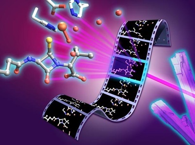
Artistic concept of the ‘molecular movie’ method used to deduce how penicillin is made. Credit: University of Oxford/Greg Stewart/SLAC National Accelerator Laboratory
More than 80 years after researchers at the University of Oxford turned penicillin into a lifesaving drug, a team there has now captured previously unseen stages in how nature builds the antibiotic’s core structure.
The β-lactam antibiotics, including penicillin, depend on a highly strained chemical ring that disrupts bacterial cell wall synthesis and ultimately kills the cell. Scientists have studied how nature constructs that ring for decades, but the key reaction intermediates exist for only fractions of a second and have been too difficult to observe directly. With one in six bacterial infections now resistant to antibiotics and too few new drugs in development, understanding how these molecules are built is an important step toward refilling the pipeline.
For the study, published in Nature Catalysis, researchers used X-ray free-electron lasers to watch an enzyme called isopenicillin N synthase (IPNS) convert its linear peptide substrate into the ring system of penicillin in a single step.
Rather than relying on static crystal structures, the team followed the reaction in real-time. They deposited thousands of tiny droplets containing anaerobic enzyme microcrystals onto a moving tape 2-mm wide. As the tape entered an oxygen-filled chamber, oxygen diffused into the crystals and started the reaction simultaneously across the sample. By controlling the tape’s speed, the researchers set how long each crystal reacted before an ultrafast X-ray pulse recorded an atomic-resolution snapshot. Combining thousands of those snapshots produced a frame-by-frame “molecular movie” of penicillin biosynthesis at physiological temperature and pressure.
The movie revealed several rapid stages, including a thioaldehyde intermediate formed just before the ring closes and a monocyclic β-lactam intermediate, the first ring-shaped structure on the way to the complete penicillin scaffold. The work also showed that water molecules inside the enzyme help guide the reaction, and that subtle movements throughout the enzyme work together with its chemistry to control the transformation.
“Penicillin has shaped modern medicine, but there is still much to learn about how nature builds this important antibiotic structure,” said Christopher Schofield, professor of chemistry at the University of Oxford and a senior author of the paper. “By capturing these fleeting steps, we can better understand how enzymes control complex chemistry with remarkable precision.”
The findings extend beyond antibiotics. IPNS belongs to a large family of iron-dependent oxygenase enzymes involved in human biology, including ones that let the body sense and respond to changes in oxygen availability. The insights could eventually inform enzyme engineering and catalyst design more broadly.
Looking ahead, the team hopes the atomic-level detail will open the door to engineering these enzymes.
“The ability to capture structural snapshots over milliseconds to seconds of reaction time allows us to connect enzyme motion, iron chemistry and water-mediated proton transfer,” said Patrick Rabe, first author of the paper at the University of Oxford. “By understanding this process in atomic detail, we can begin to think about engineering these enzymes to produce new or improved antibiotic scaffolds.”