Featured Article
Specialization and miniaturization make Raman increasingly sensitive and portable
A sample’s Raman spectrum provides information about molecular vibrations that can identify and quantify the chemicals in a material. The technology requires little, if any, sample preparation, and is used by scientists, engineers and clinicians for many purposes. According to Nick Barnett, business development manager at the Ocean Optics regional office in Oxford, U.K., the most interesting trends in Raman spectroscopy are “cancer detection, bacterial identification, process monitoring and materials science research.”
Recently, Toronto-based Tornado Spectral Systems rolled out the HyperFlux P.R.O. Plus spectrometer. It uses a proprietary high-throughput virtual slit that—according to Edward M. Albe, vice president of marketing and strategic partnerships—“allows more than 95% of the photons to go to the detector.” He adds, “HyperFlux spectrometers use a comparatively large input aperture, for maximum throughput combined with specially configured mirrors, lenses and other elements.” In combination, Albe explains, these components “compress, reformat and then expand the light beam, narrowing the input aperture along the dispersion axis while preserving total flux.” As part of the product launch, the company stated that the instrument “can acquire chemical signatures of interrogated samples with the speed and accuracy required to perform many quality and safety measurements directly on the production line.”
To provide many options on one device, some companies design particularly flexible Raman spectrometers. Last April, at the SPIE Defense Security Sensing Conference in Baltimore, Maryland, BaySpec (San Jose, Calif.) featured the portable Agility Raman spectrometer with a dual-band light source. Accessories for liquids, powders, thin slides and other sample forms are available. Its various configurations make the device applicable to many areas, the company says, including chemical-biological-explosive (CBE) detection, forensics and R&D.
Revving up Raman
Traditional approaches to Raman spectroscopy can pick up signals from the container holding the sample, which limits it to surface measurements or samples in transparent containers. According to Barnett, spatially offset Raman spectroscopy (SORS) uses two spectra taken from different positions, which leads to variations in the amount of signal from the container and the sample, making it possible to reduce the container signal in the final spectrum. SORS is known as through-barrier Raman, and it works with samples in a wide range of containers. The RapID from U.K.-based Cobalt can record Raman spectra through “opaque and colored sacks, tubs and bottles,” according to the company website.
Another advance in technology is surface-enhanced Raman spectroscopy (SERS), in which a sample absorbed in a noble-metal surface, such as gold nanoparticles, can produce a larger Raman signal. Combining these particles in a three-dimensional structure can increase the Raman signal by orders of magnitude. Gold SERS substrates from Ocean Optics make the technique easier and repeatable. These substrates can be used in many applications, such as the detection of trace levels of explosives or narcotics, as well as screening foods for additives, like melamine, or contaminants, like pesticides. Scientists also use SERS to identify biological components, including bacteria, DNA and proteins.
One challenge with SERS substrates is that the nanoparticles are not necessarily evenly distributed on the substrate. In this case, a tightly focused, static laser may miss the SERS hotspots on the sample. Raster orbital scanning solves this problem, because it uses the tightly focused laser beam to sample the substrate at many points over a much larger area. The IDRaman reader from Ocean Optics uses this technique.
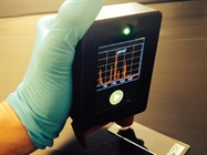
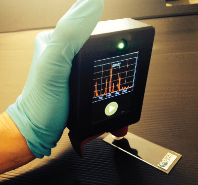 Miniaturization makes Raman spectroscopy more mobile than ever. (Image courtesy of Ocean Optics.)
SERS combined with a scanning probe microscope (SPM)—which is one kind of atomic force microscope (AFM)—makes tip-enhanced Raman spectroscopy (TERS). Coating the tip of the SPM with a metal or nanoparticles provides SERS that is limited to the area around the tip. That creates a spatial resolution of less than 100 nanometers. HORIBA (Edison, N.J.) and AIST-NT (Novato, Calif.) put together the former’s XploRA spectrometer and the latter’s SmartSPM. This system, which fits on a standard, lab-size optical table, provides chemical imaging of single molecules with a spatial resolution of just 8 nanometers. HORIBA used this platform to image single carbon nanotubes, and gathered the data in less than 10 minutes.
Other companies also offer devices that perform TERS. Renishaw (Hoffman Estates, Ill.) makes a combined Raman-SPM/AFM platform, which includes an option for TERS. Bruker (Billerica, Mass.) makes TERS-AFM probes for its Innova-IRIS, which combines AFM and Raman imaging. As the commercial options increase, more scientists will explore TERS.
This technology already interests many scientists. In a 2014 article in the Journal of Physical Chemistry Letters, researchers from Northwestern University (Evanston, Ill.) said that TERS “has experienced tremendous growth in the last five years.” Specifically, they added, “TERS imaging has provided invaluable insight into the spatial distribution and properties of chemical species on a surface with spatial resolution that is otherwise unattainable by any other analytical method.”
Like many other forms of technology, devices that perform Raman spectroscopy keep getting smaller, such as the handheld IDRaman mini 2.0 from Ocean Optics, which is only about one-tenth the size of the smallest IDRaman reader. The BRAVO handheld Raman spectrometer from Bruker is well-suited for identifying raw materials. Urban Faeh, president of Bruker Optics, expects the instrument to be used in research and quality control.
Clinical applications of Raman
In 2015, scientists at the Dublin Institute of Technology in Ireland described Raman spectroscopy’s use in screening for and diagnosing cervical cancer. In their article in Analytical and Bioanalytical Chemistry, they wrote, “There is an unmet clinical need for new methods to aid clinicians in the early detection of cervical pre-cancer.” The researchers pointed out that Raman spectroscopy detects subtle biochemical changes that arise from cervical cancer even in its early stage. The authors added that the technique applies to more than cervical cancer: “Over the past 15 years, there have been numerous reports revealing the potential of Raman spectroscopy together with multivariate statistical analysis for the detection of a variety of cancers.” Few areas of medicine strive to reduce detection limits any more than oncology, and Raman spectroscopy could add a new approach.
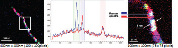 Tip-enhanced Raman spectroscopy (TERS) mapping of a single carbon nanotube showing an optical spatial resolution down to 8 nm; total map acquisition time <10 minutes (Image courtesy of HORIBA.)
It can also battle bacteria. In a 2015 article in the Annals of the New York Academy of Science, researchers from Case Western Reserve (Cleveland, Ohio) described use of the technology to discover new antibiotics and resistance to existing ones. They explained that a drug can be applied to bacterial cells, which are then frozen and freeze-dried. Then, Raman spectroscopy can quantify the number of molecules of the drug in each cell and reveal details of drug–target interactions. The researchers tested this method on two classes of drugs and various forms of gram-positive and gram-negative bacteria. They concluded, “The advantages of the present protocol are that it does not use labels and it can measure the kinetics of cell-compound uptake on the time scale of minutes.”
Raman spectroscopy provides identification and quantification for a broad collection of chemicals in an array of fields, from basic research to industry. Companies build platforms for very specific tasks or more general-purpose use, and miniaturization makes it possible to hand-carry the technology. Those are some good, good, good, good vibrations.
Mike May is a freelance writer and editor living in Ohio. He can be reached at [email protected].