Featured Article

Seeing the compounds and their distribution in samples
Looking at a slice of tissue under a microscope reveals structures, especially ones marked with dyes or other indicators, but what else is there? The more slices that scientists study, the more they wonder what makes up the structures. You can find out with imaging mass spectrometry (IMS)—a technology also known as MS imaging.
In matrix-assisted laser desorption ionization (MALDI) IMS, an ordinary tissue section is processed as usual for MALDI MS. Then, it is raster-scanned with a laser to generate a spatial map of MS data. The same slide is then stained for microscopy and imaged. The MS and imaging data sets can be combined.
“IMS is different from other imaging modalities in that it is capable of mapping hundreds to thousands of compounds, label-free, from samples such as thin tissue sections,” says Shannon Cornett, market manager for life science imaging at Bruker Daltonics (Billerica, Mass.). “In contrast to other mass spectrometry techniques, IMS is unique in that it produces spatial maps of each compound detected that shows where it is localized within the sample along with relative abundance.”
Some facilities focus on expanding the use of IMS. “The National Research Resource for Imaging Mass Spectrometry was established in 2011 at Vanderbilt University within the mass spectrometry research center through funding from the National Institute of General Medical Sciences,” says Danielle Gutierrez, project and communications manager at the center (Nashville, Tenn.). “The resource is directed by Richard Caprioli, who pioneered the development of IMS beginning in 1997.” He adds, “A driving goal of the resource is to advance imaging capabilities for the application of IMS to important biological questions and for implementation of the technology by nonexpert users within the scientific community.”
IMS can be applied to many research projects. At Vanderbilt, says Gutierrez, “The majority of imaging projects within the resource address biological and clinical issues for which the application of IMS can lead to a deeper understanding about the molecular basis of disease.” Other scientists around the world also use IMS, and a team of Japanese scientists used it to study the differentiation of stem cells.1
The use of IMS is only beginning, but some of the results already let us explore areas that we could not imagine just a few years ago. Even now, a search of “imaging mass spectrometry” turned up more than 800 articles on PubMed. That’s already a pretty good start at using IMS in research.
Clinical opportunities
“Over the past few years, two applications have driven interest and development of MALDI imaging,” Cornett says. The first is biomarker discovery within the larger scope of clinical research. “In a discovery study, cohorts of samples are analyzed by MALDI imaging and the molecular fingerprints from different regions are examined for molecular changes associated with each,” Cornett explains. “Clinical samples are analyzed by MALDI imaging in order to gain a better understanding of biochemical processes associated with disease development as well as to identify potential molecular markers that may be diagnostic of disease state, a positive or negative response to treatment, etcetera.” However, this kind of research has also been used to study nonclinical samples, including animals, insects, microorganisms and plants.
The second application driving IMS, according to Cornett, is “the preclinical imaging of dosed therapeutics and their metabolites in support of drug development. … Compared to traditional autoradiographic imaging, MALDI imaging—being mass spectrometry based—can differentiate a parent drug molecule from metabolites and the hundreds of endogenous metabolites present in tissue.” He adds, “Combine this with the lower cost of label-free imaging and the ability to correlate the hundreds of molecular changes with histopathology and one can easily see how this can benefit pharmaceutical companies.”
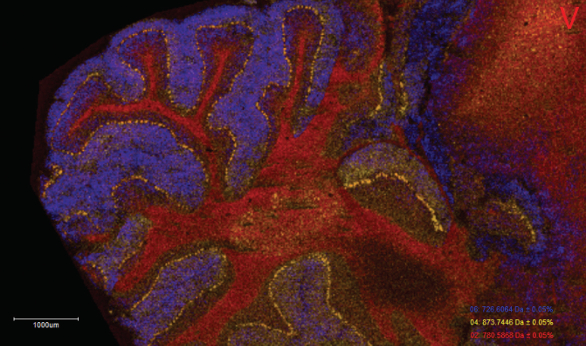 Figure 1 – This high-resolution MALDI image shows three selected phospholipid ions in transverse section of rat brain cerebellum. (Image courtesy of Bruker Daltonics.)
In addition, IMS has the potential to reveal molecular markers that may be predictive of adverse events in early stages of drug development and may help scientists better understand the biological origin of an event. “The economic advantages to earlier termination of a test article within the development pipeline can be enormous,” Cornett says.
A group of German scientists agrees that IMS offers significant benefits, and they wrote: “Since MALDI IMS enables the assessment of spatial molecular arrangements in tissue sections, it goes far beyond microscopy in providing hundreds of different molecular images from a single scan without the need of target-specific reagents. Thus, this technology has the potential to uncover new markers for diagnostic purposes or markers that correlate with disease severity as well as prognosis and therapeutic response.”2
Some scientists already use IMS in advanced forms of therapy, such as immunotherapy. Yasuhiro Matsumura of Japan’s National Cancer Center and his colleagues used IMS to track the distribution of antibody-drug conjugates (ADCs) in tumors, and concluded that IMS “is a useful tool to assess ADCs and facilitate the optimization of ADC design.”3
The clinical applications go beyond developing drugs. Richard Zare of Stanford University (Calif.) and his colleagues used desorption electrospray ionization-mass spectrometry imaging (DESI-MSI) plus a statistical technique called LASSO (least absolute shrinkage and selection operator) to “diagnose pancreatic tissue sections and prospectively evaluate surgical resection margins from pancreatic cancer surgery,” and reported that the results “provide evidence that the molecular information obtained by DESI-MSI/LASSO from pancreatic tissue samples has the potential to transform the evaluation of surgical specimens.”4
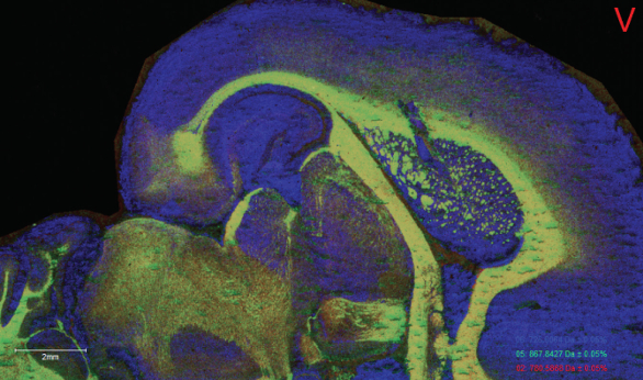 Figure 2 – MALDI imaging can explore samples, such as this rat brain, in various ways to give scientists more information about the components in a structure. (Image courtesy of Bruker Daltonics.)
Commercial creations
Scientists interested in using IMS have various options from which to choose. The iMScope TRIO imaging mass microscope from Shimadzu (Columbia, Md.) comes with software that provides a variety of analytical approaches, including principal components analysis and hierarchical cluster analysis.
Scientists can also visit the MS Imaging website (http://ms-imaging.org/wp/) to download BioMap, IMS analysis software developed by Novartis (Basel, Switzerland).
Bruker offers complete IMS systems designed to address specific applications. “Our rapifleX MALDI TOF/TOF [time-of-flight] system was designed specifically for the high sample throughput and high image resolution necessary for statistics-based discovery workflows,” Cornett says. “Bruker’s solariX XR and the newer 2XR systems are ideal platforms for imaging of therapeutics and are already deployed at many of the top 25 pharmaceutical companies.”
The best platform to use depends on the application. “The required technical features of an IMS platform will vary depending on the biological question,” Gutierrez explains. “While it is desirable for factors such as spatial resolution, mass resolution and mass accuracy to be high, there are trade-offs that come with increasing these parameters, such as the loss of sensitivity, long acquisition times and large data file sizes—for example, hundreds of gigabytes.”
Those gigabytes of information change what a scientist can see on a slide. The view changes to collections of compounds, revealing a deeper look at what makes biological things work.
References
- Shimuzu, Y.; Satou, M. et al. Matrix-assisted laser desorption/ionization imaging mass spectrometry reveals changes of phospholipid distribution in induced pluripotent stem cell colony differentiation. Anal. Bioanal. Chem. 2016; doi: 10.1007/s00216-016-0015-x.
- Schwamborn, K.; Kriegsmann. M. et al. MALDI imaging mass spectrometry—from bench to bedside. Biochim. Biophys. Acta 2016; doi: 10.1016/j.bbapap.2016.10.014.
- Fujiwara, Y.; Furuta, M. et al. Imaging mass spectrometry for the precise design of antibody-drug conjugates. Scientific Reports 2016; doi:10.1038/srep24954.
- Eberlin, L.S.; Margulis, K. et al. Pancreatic cancer surgical resection margins: molecular assessment by mass spectrometry imaging. PLoS 2016; doi: 10.1371/journal.pmed.1002108.
Mike May is a freelance writer and editor living in Florida. He can be reached at [email protected].