Cochlear amplification, a process that aids in the sensitive hearing of humans and other mammals, is thought to rely on the behavior of the motor protein prestin, which is found in the outer hair cells (OHCs) of the inner ear. Although prestin is known to consist of 744 amino acids, its three-dimensional structure within the OHC membrane has yet to be fully elucidated, and protein is also difficult to isolate from the other proteins in the membrane. Researchers at Kanazawa University have now successfully isolated and uncovered some details about the structure of prestin using single molecule force spectroscopy (SMFS) and biotin-streptavidin labeling.
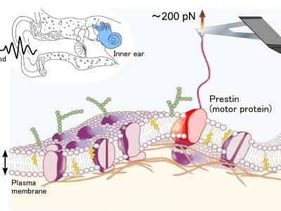
The experiment involved pulling prestin molecules from the membrane using a streptavidin-coated atomic force microscope (AFM) cantilever, which binds to the biotin-labeled prestin. As the protein is pulled through the membrane, force-extension curves are acquired indicating the amount of force needed to extract the molecule. The resulting curves had a saw-toothed pattern, as the force needed for extraction increased sharply at points where the protein was unfolding. This method allowed the researchers to identify separate domains within the protein’s structure. The researchers’ work was published in the Journal of Biomechanical Science and Engineering.
“Our results suggest that prestin has 12 transmembrane domains, which supports the predictions of a previous model,” said first author Michio Murakoshi. “Our findings demonstrate the potential of our biotin-streptavidin method for investigating proteins using SMFS, and provide important information on the response of OHC membrane proteins, contributing to a better understanding of cochlear amplification.”
The researchers noted the value of using biotin-labeling in this experiment due to biotin’s small size, strong interaction with streptavidin, and the fact that it does not have any significant effect on the behavior of the target molecule.
Photo: Force spectroscopy of prestin, the motor protein in the inner ear. Credit: Kanazawa University